


This disease has been poorly studied, although several thousand observations with diagnosis and subsequent treatment have been described.
Many variety and not -the specificity of the clinical picture of the varicose veins of the pelvis lead to gross errors from the diagnosis, which in the future affects the consequences.
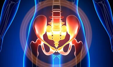
Characteristics of varicose veins of small pelvis
The pelvic veins are several times longer than the arteries, which determines their large capacity.This is due to the phylogenesis of the vascular system of the pelvic region.The pelvic veins have high adaptation capabilities and are potentially prone to restructuring, which contributes to the formation of a densely intertwined network.
The speed and direction of the blood flow are regulated by valves that are controlled by complex humoral mechanisms.The valves balance the pressure in different parts of the venous network.
When the valves cease to perform their functions, blood stagnation develops, this leads to the pathology of the blood vessels and the formation of varicose veins.The uniqueness of the pelvis of the pelvis lies in the fact that the wide ties of the uterus, which keep the lumen of the vessel wide, can also narrow it, causing pathology.
Reasons
Pathological dilation of pelvic veins may be due to the following reasons:
- Disturbance of blood flow paths;
- Venous barrel prison;
- Compression of collateral trunks from the altered position of the uterus, for example in retroflexia;
- Damage to the ovaries of the valve (congenital or acquired);
- Obstructive post -fluid syndrome;
- Connective tissue pathology;
- Arteriovenous angiodisplasia;
- Long -term meeting, heavy physical labor;
- Varicose veins of the lower limbs;
- Pregnancy (3 or more) and birth (2 or more);
- Diseases of the female genital area (chronic salpingophoritis, ovarian tumors, uterine fibroids and genital endometriosis);
- The adhesive process of the pelvic organs;
- Obesity.
Grade classification
In terms of enlarged vein, the following degrees are distinguished:
- up to 0.5 cm, Tirbushon vascular impact;
- 0.6-1 cm;
- More than 1 cm.
Options for the course of the disease
- varicose veins of the perineum and vaginal lobby;
- Venous adult pelvic syndrome;
Symptoms
- The most common pain in the lower abdomen, perineum after long static and dynamic surge.The pain is exacerbated in the second phase of the cycle, after hypothermia, fatigue, stress, exacerbation of various diseases.
- The feeling is "not calm", pain with and after it.
- Dysmenorrhea - menstruation disorders, including pain syndrome.
- Secretion, more than normal, bile of genital tract.
- Blood stagnation leads to infertility, inability, termination of pregnancy.
- Urination disorders as a result of the expansion of the bladder veins.
Diagnostics
The diagnosis of the disease alone from complaints is successful in only 10 % of cases.
The palpation of the inner walls of the pelvis allows to feel elongated seals and nodes of the veins.When examined in the mirrors, the cyanosis of the mucosa of the vagina is visible.
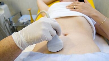
The selection procedure is an ultrasound examination with color Doppler mapping, which allows you to identify not only the variations of varicose veins of ovarian veins, but also venous thrombosis, post -clomboflebitic occlusion.Ultrasound shows "worms", the structure without reflection of the signal located on the lateral surface of the uterus.
The effect of Doppler examination is based on "toning" in blue and red, venous and arterial blood, corresponding.
The ultrasound examination apparatus, using a special program, recognizes blood movement from the sensor, and in the other direction it calculates the blood flow and the type of vessel.
But the exact definition of the vein is or the artery remains behind the doctor.The Doppler method works in almost all cases, our body dictates exceptions to the rules, since the blood that flows from the heart is not always arterial and vice versa.
Thus, an ultrasound doctor sees an arterial or venous vessel, its size, the rate of blood flow in it, and many indicators that an ordinary person does not need, but plays an important role in diagnosis.To do this, use transabdominal and transvaginal sensors.
In 5.7% of cases, the disease is recognized accidentally during screening.Usually the diameter of Vena Ovaric is 0.4 cm.
CT and MRI have great accuracy.Using these methods, you can find varicose veins in uterine ligaments, ovaries and around these organs.It is possible to determine the accompanying pathology.
A very reliable method is a phlebographic study.
The contrast is carried out at the height of the Valsalva sample, against blood flow.This allows you to see the valve damage.
Retroneoscopy is also used on the left, renal phlebography, super -follow -up phlebavroscopy and phleburialography on both sides.These methods allow you to determine the hemodam and the anatomical changes in the kidney veins and the places to fall into them on gonadal veins.
Super -Spent phlebovarioscopy is performed by catheterization of gonadal veins through a contrasting femur or subclavian vein, with subsequent introduction of contrast.
The greater part of the blood from the varicose horn plexus is discarded through the ovarian vein.But in conditions of hypertension, it occurs through unstated uterine veins in the inner iliac vein.The plexus of the veins, through which the leakage may occur, includes sacral and plexus of the bladder.
In the left phlebography, 3 stages of venous stagnation are distinguished in the cluster split of the left ovary:
- There is no leakage from the left ovary plexus or is carried out on an additional short path.
- There is an extra long road.
- There are two additional paths of leakage or one additional and ancillary.
At 2 and 3 stages varicose veins of the cluster plexus of the right ovary are formed.
Laparoscopy is used for differential diagnosis.The pathologically confused veins are in the ovary, in the direction of round and wide ligaments.They look like large cyanotic conglomerates with a thin and tense wall.
The complexity of the diagnosis lies in the fact that the disease is often hidden behind the signs of the inflammatory process, is characterized by clinical manifestations, masked in endometriosis, prolapse of internal organs, postoperative neuropathies and very extragenic conditions.
Treatment
The main purpose of treatment is to eliminate the reflux in the veins.Conservative treatment is used in the initial stages of the disease.In the late stages of the disease, the treatment of choice is surgery.
Conservative treatment

It is to normalize the tone of the veins, improve hemodynamics and trophic processes.
Symptomatic treatment to eliminate individual symptoms.Non -steroidal anti -inflammatory pain, bleeding - hemostatic therapy.
The main medicines for conservative treatment are venotonic drugs and anti -platlates.
Phlebotonics - Improve the tone of the vascular wall and enhance blood flow.In this disease, it is better to consult a gynecologist for certain medicines.
An important method is physiotherapy exercises.
Surgical treatment
- Revival of varicose veins.
- Gonado-Cavalry Shunning.
- Sclerosis for laparoscopy.
- Occrames of ovarian veins using X-ray-endovascular methods.
Folk remedies
Since the main thing in the onset of the disease is the weakness of the valve apparatus, all folk remedies used to expand the varicose veins of the lower limbs are also used for this pathology.
The most commonly used: ordinary hazelnut, hops, nettle, horse chestnut, dandelion root, tea sponge, willow, oak, St. John St. John, series, pollen and many other plants.
It is effective: oak bath treatment, chestnut, willow, chamomile, pharmacy, herbs of drying, St. John's wort.
Prevention
- The first thing you need to do if there are complaints, forecasts or diseases listed above - contact a gynecologist.
- It is necessary to normalize the operating mode and to rest, to try not to stay upright for a long time, physical tension.
- Do exercises for the "Pedal", "Stack of Standup", "Legs"
- Stick to the diet: Eat foods high in vitamins E, R, C, try to eat only white meat, less fat, replace it with fruits, vegetables, cereals.
- Drink enough liquid, but not less than 1.5 liters per day.
- Get rid of excess weight, bad habits.
- Consult your doctor about wearing compression linen, this will improve blood flow from the lower limbs, thus there will be less stagnation in the pelvis.
- Avoid bathrooms, saunas, steam rooms, hot baths.
In order not to become ill with such a difficult disease, it is necessary to follow the preventive recommendations listed above.Treat your health as the most valuable in life.
For the sorts of suspicious symptoms that you cannot get rid of for several days, you should consult a doctor.He should provide you with highly qualified help and save you from suffering.